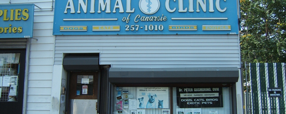
Call Us Today! (718) 257-1010 |

Animal Clinic of Canarsie
  • Home
  • Meet our Team
  • Services Offered
  • Links
  • Resources
    • Education
      • Canine Articles
      • Feline Articles
  • Online Pharmacy
  • Emergencies
  • Contact Us
  • Home
  • Meet our Team
  • Services Offered
  • Links
  • Resources
    • Education
      • Canine Articles
      • Feline Articles
  • Online Pharmacy
  • Emergencies
  • Contact Us

Pet Radiology

In Blog
AdobeStock_89700933-300×198

Have you ever wondered how we take animal x-rays and perform other types of diagnostic imaging on dogs and cats?  Well, the equipment is much the same as what is used on humans. 

When x-rays are used, the animal is placed on a special table and an x-ray machine is used to take the images.  The machine releases an x-ray beam that passes through the part of the animal that is under the beam.  Different types of tissues absorb x-rays to different extents, and this is how an image is created.  Some hospitals have digital x-ray processing, where the picture comes up on the computer screen, and some hospitals have manual x-ray processing, where film is used and processed using another machine.

The main difference between human and animal x-rays is that we can’t ask our patients to ‘stay still’ for us to take the x-ray!  In order for animals to be correctly positioned, they either need to be held in position by gentle restraining devices or veterinary staff, sedated or sometimes anesthetized.

An ultrasound  for dogs or cats is another imaging modality that we like to use to assess our patients.  In this technique, an ultrasound machine with a probe is used.  The probe emits ultrasound waves that provide a black and white picture of the organs on the screen.  Ultrasound is most commonly used to assess the abdomen or the heart, but other areas can also be imaged using this technique.  Ultrasound of the heart is called an echocardiogram.  Most animals can receive an ultrasound while being physically restrained by veterinary staff, however some animals may need sedation.

Advanced imaging refers to Magnetic Resonance Imaging (MRI) or Computed Tomography (CT).  These techniques are particularly useful to assess the spine, brain and lungs, but are commonly recommended for the assessment of many other areas.  General anesthetic or heavy sedation is required to carry out these tests.  MRI and CT are only available in larger hospitals.

Did you like this article? Share it with your friends!

The post Pet Radiology appeared first on Pet Assure Blog.

Source: Pet Assure Blog

Previous StoryBleeding and Other Cat Paw Disorders
Next StoryUnderstanding Your Dogs Sleeping Pattern

Related Articles

  • dog
    Three Natural Ways to Get Rid of Dog Tartar
  • Rottweiler-Mouth
    Braces for Dogs

Recent Posts

  • Three Natural Ways to Get Rid of Dog Tartar
  • Braces for Dogs
  • Cats and Christmas Trees
  • My, What Shiny Teeth You Have
  • Do You Pay Enough Attention to Your Pets?

Recent Comments

    Archives

    • March 2020

    Categories

    • Blog

    Meta

    • Log in
    • Entries feed
    • Comments feed
    • WordPress.org

    WELCOME!

    We offer a variety of wellness and vaccination packages which will save you 25% compared to our regular fees.

    RECENT POSTS

    • Three Natural Ways to Get Rid of Dog Tartar
    • Braces for Dogs
    • Cats and Christmas Trees

    BUSINESS HOURS

    We are open Wednesday through Saturday by appointment only. We're closed Sunday, Monday and Tuesday.

    Animal Clinic of Canarsie
    • Home
    • Meet our Team
    • Services Offered
    • Links
    • Online Pharmacy
    • Emergencies
    • Contact Us

    Copyright | Powered by DVM Galaxy